Information
Oct 23, 2018
Research on Automatic Detection of Cerebral Aneurysms on MRA Using Deep Learning
In this study, we developed a deep learning-based tool that identified cerebral aneurysms from Magnetic Resonance Angiography (MRA) data. The paper presents the steps we took to develop this tool using MRA collected from multiple institutions, and to verify its detection sensitivity and clinical utility.
Paper
Deep Learning for MR Angiography: Automated Detection of Cerebral Aneurysms
Radiology
https://doi.org/10.1148/radiol.2018180901
Author's Comments
This was our team's first doctoral dissertation, and we received significant support through collaboration with LPixel, a company that possessed cutting-edge medical image analysis technology in Japan at the time. I believe it was very meaningful to be approached during the early adoption of deep learning in the medical field and to develop an algorithm targeting cerebral aneurysms. Furthermore, the model we developed achieved approval as Japan's first deep learning-based medical device and was selected as "The Best of Radiology" at RSNA. Such recognition was possible only because of the cooperation and support from many people, especially LPixel, to whom we are particularly grateful.
Paper Overview
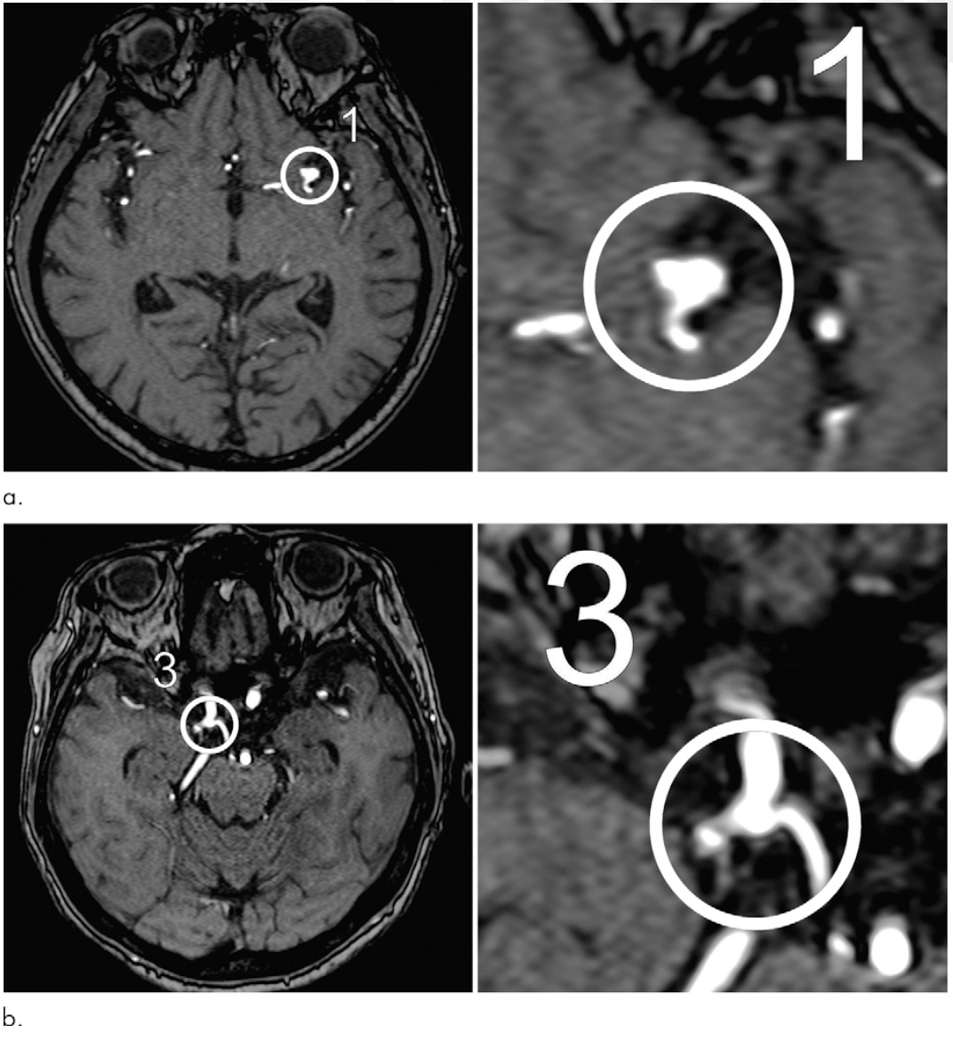
The aim of the research was to develop an algorithm to assist in preventing overlooked cerebral aneurysms using head MRA (specifically the TOF method). We divided images collected from four different medical institutions into training and validation sets, aiming to demonstrate stable analytical performance even in environments with different image sizes and imaging conditions. We used ResNet-18 as the base deep learning model, and trained it to extract candidate points for cerebral aneurysms from each patient's MRA image and to distinguish whether the candidate was a true aneurysm. As it was originally designed with "prevention of oversight" in mind, we adopted an approach to maximize sensitivity as much as possible.
Paper Details
We collected more than a thousand MRA imaging cases, of which 683 cases were used as training data for the algorithm. When validating the completed model with internal as well as external test datasets collected from different institutions, it detected cerebral aneurysms with sensitivities of 91% and 93%, respectively. Small aneurysms (less than 3mm in size) were detected at an average rate of about 89%. Additionally, when this model was added as an aid to support conventional reading, the detection rate increased by 4.8% for the internal dataset and 13% for the external dataset. Furthermore, multiple new aneurysms were found in relatively low-incidence locations such as the vertebral artery and basilar artery, which had not received much attention previously. We believe our efforts to ensure the model could adapt to different imaging conditions and devices were successful. Through these research results, we feel we've demonstrated the potential to improve reading efficiency and reduce oversights. In the future, we plan to expand the scope of this technique, conduct verification under more diverse conditions, and aim to improve accuracy in clinical practice.