Information
Mar 30, 2021
Development and Evaluation of Cerebral Angiography Method without Misregistration Using Deep Learning

In order to eliminate misregistration artifacts caused by patient movement, we validated a model that uses deep learning to generate images comparable to digital subtraction angiography (DSA).
Paper
Deep Learning–based Angiogram Generation Model for Cerebral Angiography without Misregistration Artifacts
Radiology
https://doi.org/10.1148/radiol.2021203692
Author's Comments
We considered how AI could be utilized in the field of IVR (Interventional Radiology) and, despite having a limited number of cases from a single institution, we achieved publication by pursuing originality in our concept. During our research, we examined in detail the mechanism of misregistration artifacts frequently seen in cerebral angiography and identified the problem where bones and devices in the background appear as residual images due to slight patient movement. We thought this challenge could be solved through deep learning. Although we initially had limited expertise in image processing and went through much trial and error, we feel we were able to build a model with high clinical practicality by pooling the collective wisdom of our entire laboratory.
Paper Overview
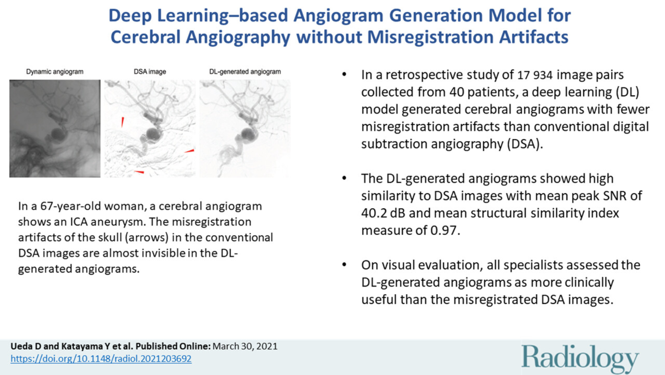
In this paper, we proposed a method to directly generate DSA, which is essential for treatment planning and during procedures for conditions such as cerebral aneurysms, using a deep learning model rather than subtraction processing with mask images. Conventional DSA can cause "misregistration artifacts" where parts of bones or devices remain visible when patients move slightly under fluoroscopy. This can make blood vessel paths unclear and, in some cases, necessitate retaking images, risking prolonged treatment time. In this study, we extracted cases from patients' past images, created training data by pairing angiography without artifacts and motion images, and trained a deep learning model to accurately extract only the vascular portions.
Paper Details
We ultimately used more than 17,000 image pairs obtained from 40 patients, dividing them into training and test data for evaluation. As a result, compared to DSA images without misregistration, the vessel images generated by the deep learning model showed high values with an average peak signal-to-noise ratio (PSNR) of 40.2 dB and an average structural similarity (SSIM) of 0.97. This means that images with comparable quality were obtained while minimizing artifacts from bones or metal coils due to movement. Additionally, visual evaluation by multiple specialists determined that the images were either more viewable than conventional DSA or showed no significant difference. Although this is a single-institution study with a limited number of cases, we believe there is significant value in demonstrating the possibility of depicting cerebral vascular structures without mask images using AI. In the future, we expect this could contribute as a means to reduce the risk of retaking images and procedural delays while lessening patient burden and radiation exposure.