Information
Jul 6, 2023
Multi-Institutional Collaborative Research on AI for Estimating Cardiac Function and Valvular Disease from Chest X-rays

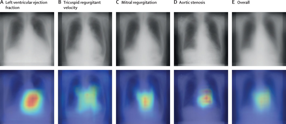
We performed a multi-institutional collaborative study demonstrating that deep learning models can estimate multiple cardiac function indicators, such as left ventricular ejection fraction (LVEF) and valvular diseases, with high accuracy from chest X-ray images.
Paper
Artificial intelligence-based model to classify cardiac functions from chest radiographs: a multi-institutional, retrospective model development and validation study
The Lancet Digital Health
https://doi.org/10.1016/S2589-7500(23)00107-3
Author's Comments
We began our research with the idea that the cardiac condition could be somewhat understood from the CTR (cardiothoracic ratio) on chest X-rays, but initially, it was a small-scale attempt to estimate only LVEF (left ventricular ejection fraction) at a single institution. Subsequently, we expanded our analysis to include cardiac valvular diseases and developed an approach to enhance accuracy and reproducibility by using data from multiple institutions. In this process, we set a goal to submit to The Lancet Digital Health, which took about five years for preparation, data collection, model construction, and refinement. While there were technical challenges and establishing inter-institutional cooperation was time-consuming, I feel we were finally able to bring it to fruition with the support of many people.
Paper Overview
We created and validated a deep learning model by combining chest X-rays collected from multiple institutions with corresponding echocardiography results. Ultimately, we obtained a total of 22,551 X-ray images from four medical institutions, comprising 16,946 patients. The model was designed to not only estimate LVEF but also simultaneously classify multiple parameters obtained from echocardiography, such as the severity of cardiac valvular diseases including mitral regurgitation and aortic stenosis, tricuspid valve regurgitation, and inferior vena cava diameter. Analysis of 3,311 external validation images showed an AUC of 0.92, sensitivity of 82%, and specificity of 86% for the task of estimating whether LVEF was less than 40%. Good performance was also demonstrated for tricuspid regurgitation with an AUC of 0.92, and other valvular diseases generally achieved AUCs in the upper 0.80s.
Paper Details
An important aspect of this study was enhancing the model's versatility by collecting data from multiple institutions and pursuing estimation accuracy based on clinical realities. Since chest X-rays are one of the most frequently performed examinations globally, they could be useful for roughly understanding cardiac function in regions where specialized echocardiography is difficult to implement or in emergency situations. While images alone cannot capture the complete picture of heart disease, our AUC of 0.92 indicates significant value in being able to estimate important indicators like LVEF quickly and with a certain level of accuracy. Future challenges include application to X-rays taken in supine rather than standing positions and additional research for further accuracy improvement. This model received no development funding, and we hope to advance implementation in clinical settings through source code that will be freely available in the future. By leveraging chest X-rays as a versatile tool, we aim to open a path that leads to prompt diagnosis and treatment for more patients.