Information
Aug 1, 2023
Creation and Validation of AI for Synthesizing Methionine PET from Contrast-Enhanced MRI

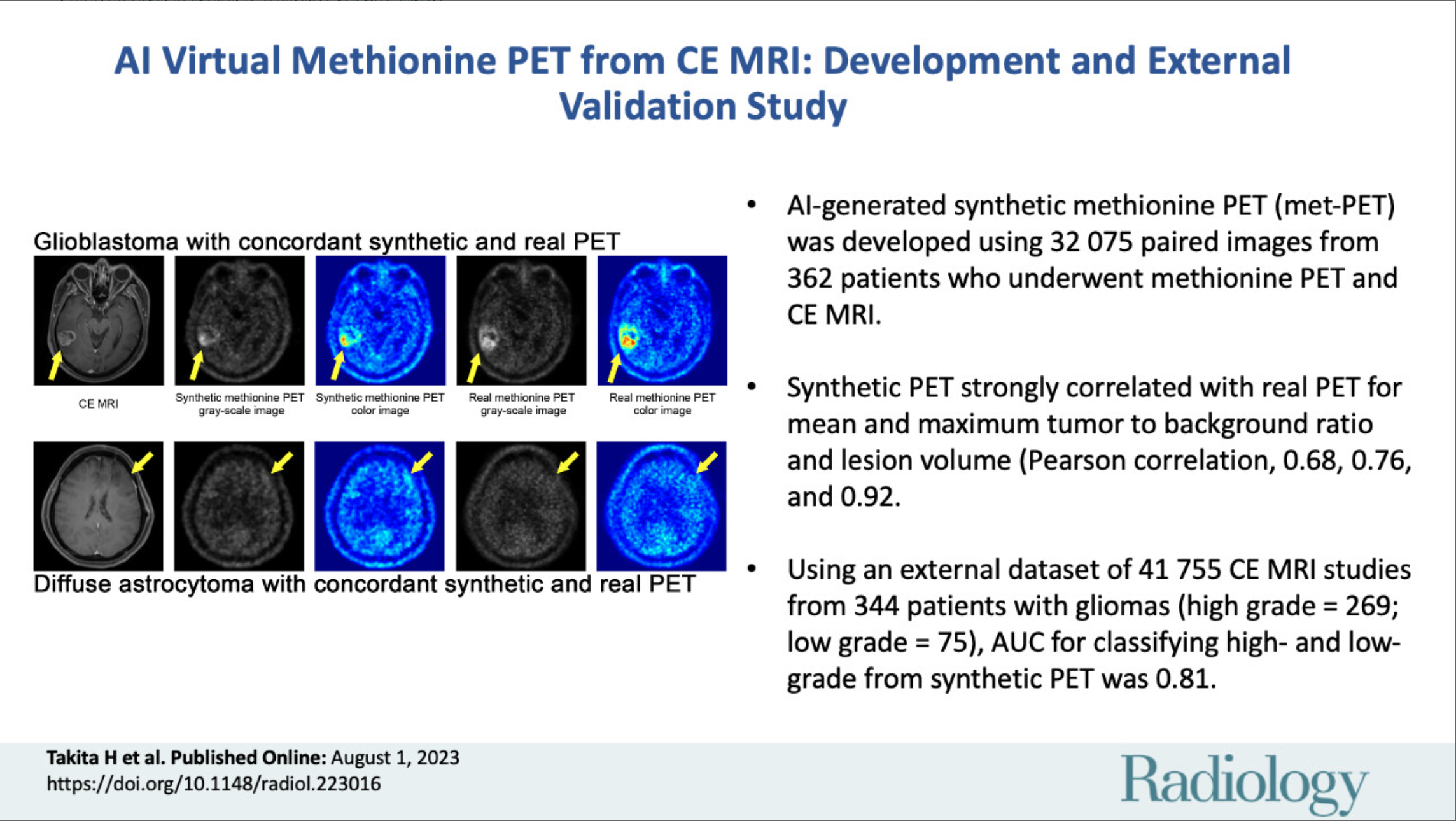
We validated the utility of a method that synthesizes methionine PET images using artificial intelligence based on contrast-enhanced MRI to evaluate malignancy and prognosis.
Paper
AI-based Virtual Synthesis of Methionine PET from Contrast-enhanced MRI: Development and External Validation Study
Radiology
https://doi.org/10.1148/radiol.223016
Author's Comments
We have been continuously exploring ways to make methionine PET, which is particularly useful for brain tumor diagnosis and treatment planning, available to more institutions and patients. However, methionine PET is difficult to implement due to its use of radioisotopes, and nuclear medicine facilities and equipment are limited. Therefore, we focused on whether AI-based image conversion based on contrast-enhanced MRI could provide information similar to actual PET. In this study, we combined international open data and multi-institutional datasets to quantitatively validate the malignancy and prognosis prediction performance. We also compiled numerous experiments into one paper to advance AI applications in the field of nuclear medicine, so I hope you will also review the supplementary materials. The fact that the first author of this work, Dr. Hiroaki Takita, received the Kato Award from the Neuroradiology Society has been a great encouragement for us.
Paper Overview
This study generated synthetic methionine PET images using contrast-enhanced MRI images as input and evaluated their accuracy and clinical utility. First, we performed training and validation using data from 362 patients obtained from our university hospital, followed by using cases from 344 patients from international institutions as external test cases. As a result, we confirmed statistically high agreement between synthetic images and actual methionine PET, with correlation coefficients of 0.68 for maximum tumor-to-background ratio (TBRmax) and 0.92 for lesion volume. Furthermore, in external test data, the differentiation between high and low malignancy likelihood using TBRmax from synthetic PET images showed an AUC of 0.81, and significant differences were observed in 2-year survival rates between low-risk groups (71% survival) and high-risk groups (27% survival).
Paper Details
We focused on the possibility that the "tumor metabolic activity" supporting the high diagnostic power of methionine PET and features such as "tumor vascular permeability" captured by contrast-enhanced MRI might have certain correlations. We incorporated a generative adversarial network (pix2pix model) for image conversion, implemented techniques to capture the three-dimensional morphology of brain tumors, and performed data sampling that included not only lesion areas but the entire brain. In internal tests, we obtained results close to measured PET not only for TBRmax and TBRmean (correlation coefficient 0.76) but also for lesion volume. Furthermore, in validation using external databases, stable values with an AUC of 0.81 were shown when discriminating between high and low malignancy likelihood using synthesized PET images, and clear differences were also observed in 2-year survival rates after treatment. While we must avoid raising excessive expectations, we believe there is significant value in demonstrating that synthetic methionine PET can potentially provide indicators generally comparable to actual measurements. However, larger-scale validation and prospective trials are necessary for clinical implementation, and we would like to continue supplementary experiments and long-term follow-up in the future.