Information
Aug 16, 2023
Clinical Application of Aging Biomarker Developed from Chest X-rays of Healthy Individuals
This research demonstrates that "apparent age" estimated by artificial intelligence based on chest X-ray images is associated with chronic diseases such as hypertension and lung diseases through the gap (difference-age) with actual age.
Paper
Chest radiography as a biomarker of ageing: artificial intelligence-based, multi-institutional model development and validation in Japan
The Lancet Healthy Longevity
https://doi.org/10.1016/S2666-7568(23)00133-2
Author's Comments
We had been thinking for some time that chest X-rays might show certain bodily changes associated with aging to some extent. While there have been previous reports of such attempts, they primarily focused on subjects with diseases, or were based on single-institution data, and we couldn't find research that created AI models using healthy individuals collected on a large scale from multiple institutions. Therefore, we collected data from healthy cohorts across multiple institutions to verify the accuracy and versatility of an age estimation model to demonstrate its usefulness. We then used the difference between the estimated age and actual age as an indicator to comprehensively examine associations with various chronic diseases. Furthermore, we utilized the Hugging Face platform to make it accessible to everyone and prepared a user-friendly GUI. We hope that this research, based on information obtained from X-rays, will serve as a foundation for diagnosing and preventing various diseases in the future.
Paper Overview
We constructed and validated the accuracy of a system that estimates age using an AI model based on chest X-ray images of health examination participants obtained from multiple facilities in Japan between 2008 and 2021. Ultimately, we analyzed over 100,000 X-ray images from approximately 70,000 individuals, and in external validation, obtained results showing a correlation coefficient of 0.95 between estimated and actual age, a mean squared error of 15.0 years, a root mean squared error of 3.8 years, and a mean absolute error of 3.0 years. Subsequently, when this model was applied to a cohort with chronic diseases collected at different facilities, we observed a trend where the greater the difference between estimated age and actual age, the higher the prevalence of various chronic diseases.
Paper Details
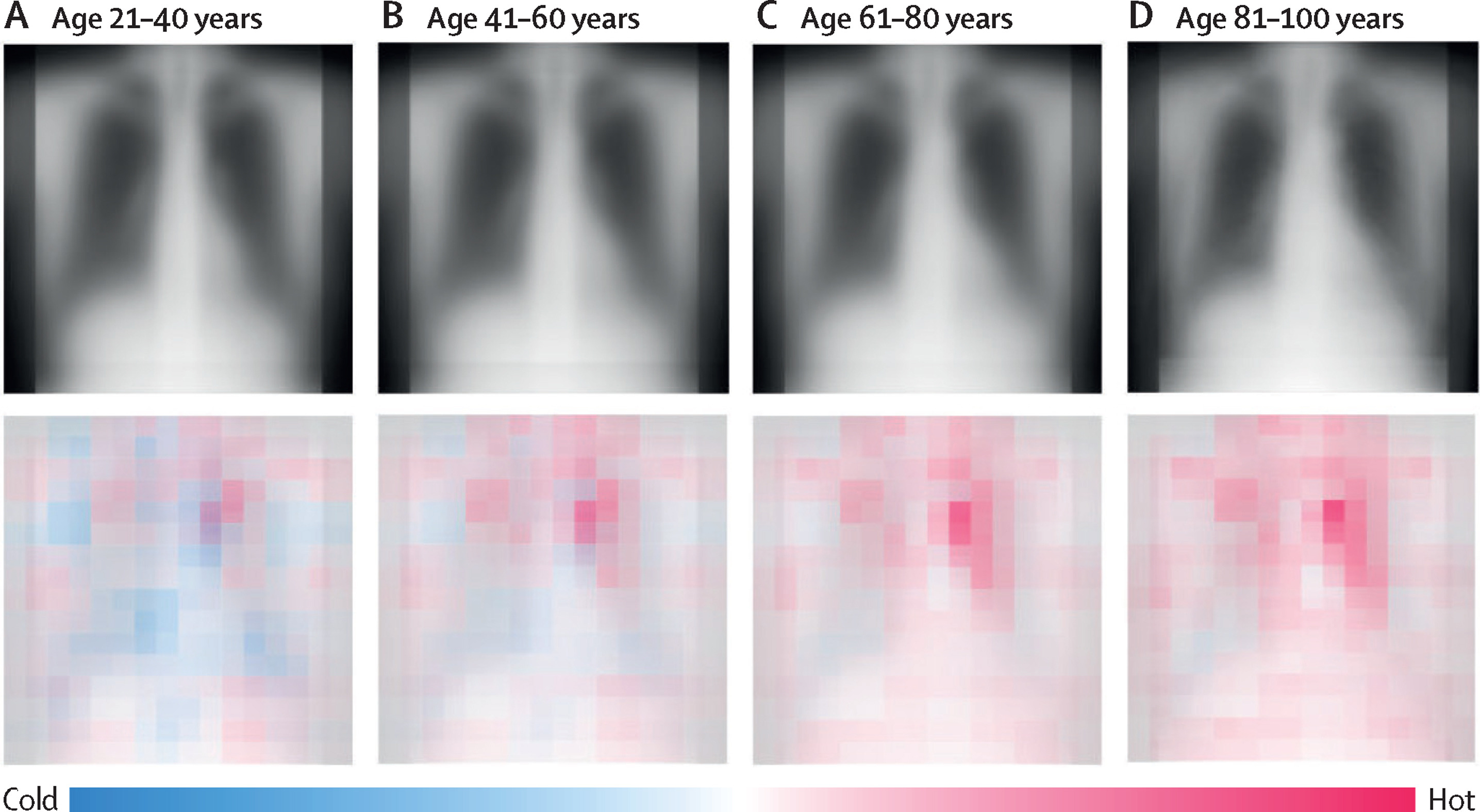
We applied the model developed from healthy cohort data to a population with diseases and verified that when estimated age was higher than actual age, there was a stronger association with chronic diseases. For example, for blood pressure abnormalities (hypertension), the odds ratio was 1.04 for each unit increase in estimated age difference, 1.05 for COPD (chronic obstructive pulmonary disease), 1.08 for interstitial lung disease, 1.05 for liver cirrhosis, and 1.03 for osteoporosis. On the other hand, the association with acute diseases was not as strong, suggesting that what the AI model captures is closer to long-term physical changes. Furthermore, when investigating which parts of the image information influenced age estimation, it was suggested that areas near the aortic arch and the lung bases served as important clues. This research indicated the potential of chest X-rays, despite being an inexpensive examination that can be performed regularly, to reflect the degree of aging. In the future, we plan to conduct comparative verification to more accurately capture biological aging and continue further validation to use this as a guideline for risk assessment, early prevention, and treatment of various chronic diseases.