Research Results
Jan 20, 2023
- Lecture / Seminar
- NEWS
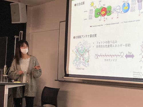
A symposium at the Research Center for Artificial Photosynthesis was held.
- Date & Time: Friday, January 20, 2023, 5pm-6pm
- Place: Sugimoto Campus, Science Building, Room E211 (Lecture Room 10)
- Speaker: Dr. Nami Yamano (Renmin University of China)
- Title of the Lecture
「脂質ナノディスクを利用した光合成アンテナ蛋白質の構造機能研究」
Structural and functional studies of photosynthetic antenna proteins using lipid nanodiscs
Ms. Yamano, who earned a Ph.D. in Chemistry from Osaka City University and is currently a postdoctoral researcher at Renmin University of China, will be giving a lecture on her recent research findings during her short-term visit to Japan. We encourage you to attend.
Caretaker: Ritsuko Fujii (ext. 3624) E-mail: ritsuko[at]omu.ac.jp (Change [at] to @)
Abstract
Photosynthesis is a process by which light energy from the sun is used to convert water and carbon dioxide into sugar. This capture of sunlight is achieved by antenna proteins embedded in the thylakoid membrane lipids, which bind chlorophyll and carotenoids as light-harvesting pigments. Interestingly, antenna proteins are not only involved in light harvesting but also play a role in dissipating excess excitation energy within the membrane in response to fluctuations in sunlight intensity. While it is known that this quenching process is caused by changes in the excited state of pigment molecules induced by protein-protein aggregation or alterations in the surrounding environment, there is a discrepancy between molecular functional analyses using isolated proteins and analyses of macroscopic samples such as leaves or thylakoid membranes. Consequently, the precise molecular mechanism of light harvesting and quenching by antenna proteins within the thylakoid membrane remains unclear.
Typically, functional analysis of photosynthetic proteins is conducted by dispersing isolated proteins in aqueous solutions containing detergent micelles. However, the high permeability of these micelles to polar molecules and ions makes it difficult to fully replicate the biological environment. Liposomes have been widely used as a system to mimic the hydrophobic membrane environment. However, it has been difficult to control random protein aggregation in liposomes, leading to heterogeneous excitation states of individual molecules. Therefore, in this study, we attempted to reconstitute antenna proteins into lipid nanodiscs, which are lipid bilayer discs. By adjusting the length of the scaffold protein, nanodiscs allow for the control of the disc size and the number of encapsulated proteins, enabling single-molecule functional analysis under lipid-like environments. Using nanosecond time-resolved absorption and fluorescence lifetime measurements, we investigated how the energy transfer and structure of pigments bound to antenna proteins differ in lipid nanodiscs compared to those in conventional detergent micelles. The results revealed that pigments in the lipid membrane tend to adopt more quenched conformations compared to those in micelles.
(Bibliography: Yamano et al., J. Phys. Chem. B, 126(14):2669-2676, 2022.)
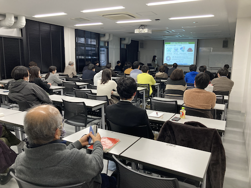
We held the meeting in person with appropriate infection prevention measures in place. Thank you for the participation and discussion of approximately 30 attendees.